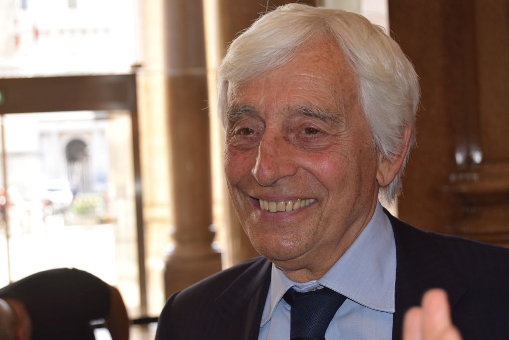
ROME (ITALPRESS) – The 126th National Congress of SIC-Italian Society of Surgery, which was held in Rome between Oct. 13 and 16 and was dedicated to “Laparoscopic Cholecystectomy: skills, prevention and multidisciplinary treatment,” closed with a speech by Professor Gianfranco Gualdi, a medical luminary and Scientific Director of the “Diagnostic Imaging” service of the Institute of Medicine and Sports Sciences of the Coni. Professor Gualdi’s talk focused precisely on the complications, risks and their possible prevention that are typical of laparoscopic cholecystectomy surgery, the minimally invasive surgical procedure for gallbladder removal that is becoming increasingly popular.In his presentation, Professor Gualdi delved into the possible complications of laparoscopic cholecystectomy with demonstrative images of fluid collections, abscesses and bleeding that can occur during or after the procedure. In addition to this, possible biliary tract resections and vascular injuries that can complicate the procedures were reviewed.In his report, the professor noted the complications that can occur after laparoscopic cholecystectomy, complications that can involve the biliary tract with the development of a biliary tract lesion of a biloma, i.e., a collection of bile, or with acute biliary obstruction or true biliary stenosis. Other complications are calcinosis that may occur in the cystic duct or dispersion of a stone intraperitoneally, or even a recurrence of calcinosis.Iatrogenic injuries affect the biliary tract and may involve hemorrhage or pseudo aneurysms i.e., unraveling of the vessel walls with bleeding, or ischemia due to vessel ligation or duodenal injury with migration of surgical clips. Similarly, recurrence of cholecystitis may occur and distant cholecyst tumor may develop if subtotal cholecystectomy is performed. In light of these complications, therefore, the use of imaging techniques for the prevention and eventual treatment of these complications is essential. In addition to this, it is important to make use of CT and MRI, which are invaluable in studying the cause of the complication with great accuracy.
– Photo: Photogram Agency –
(ITALPRESS).

